Electrical Muscle Stimulation Principles
Click HERE if you are looking to learn more about the benefits of the STIMFIT on your body.
Muscle contractions are produced and controlled by the brain by means of electrical signals transmitted through the nervous system. When an electrical signal from the brain reaches the muscle, the latter is activated into groups of “motor units”, each made up of a single neuron and of a group of associated muscle cells connected to it. This initiate a chemical reaction which causes the cells in this motor unit to contract.
The complete contraction of the muscle usually involves a number of motor units simultaneously, and its strength is directly proportional to the number of activated motor units. The gradual enrollment process of the motor units which consents to a perfectly controlled and smooth muscle contraction is called spatial summation.
Similar muscle activation’s may be obtained, without cerebral input, by means of electrical neuromuscular stimulation.
When a sufficiently intense single electrical impulse reaches the motor muscle or nerve, it causes one short single contraction of the muscle (spasm). If this single spasm is repeated and the frequency of reiteration exceeds ten spasms p.s., each following spasm is enhanced by one degree of muscle shortening caused by the preceding spasm. Such an effect is called temporal summation. The lowest stimulation frequency, where the successive contractions merge, is called tetanization frequency. Tetanization frequency usually ranges between twenty-five and fifty impulses p.s. in accordance to muscle type and is used to induce tetanic muscular contractions which are stimulated electrically.
The strength of natural muscle contractions is partially obtained by the summation of reiterative spasms which take place within the motor units (temporal summation) and partly by the total amount of recruited motor unit which carry out the contraction (spatial summation). During motor excitation electrostimulation the strength of muscular contraction peeks if tetanus stimulation frequency is reached; however it also depends on the total amount of motor unit recruited in the stimulation.
The number of motor units recruited in the stimulation is determined by the energy of the impulse supplied to the motor muscle or to the motor nerve. The latter is a function of the length of the impulse itself (impulse width) and of the power intensity during the stimulation impulse.
Generally speaking, the intensity of the stimulus supplied to the motor nerve/muscle depends on:
- Intensity of the power impulse supplied by the stimulator and determined by its control panel;
- Skin resistance and electrode-skin interface resistance: the smaller the resistance, the greater the intensity of the impulse;
- The placement of the electrode: the smaller the distance between the electrode and the motor nerve/motor muscle area, the greater the strength of contraction.
If the electrical neuromuscular stimulation is to be effective, the energy supplied to the motor nerve/motor muscle area must be regulated through the push buttons on the electrical stimulator in the best possible way.
The best effects of stimulation are obtained when the largest possible number of motor units are supplied with enough energy to overcome their activation threshold. Such energy may, however, be limited by the discomfort which may arise by stimulating the pain receptors located on the skin. An adequate setting of parameters is therefore paramount on behalf of the manufacturer together with the correct stimulation procedure adopted by the user, so as both to maximize the energy supply and avoid feelings of unease.
Muscle Structure and Physiology
Muscles are made up of muscle fibers, the extremity of which are connected to muscle tendons. When activated or stimulated by impulses from the brain, muscle fibers contract and the resulting strength of contraction is transmitted to the bones via the tendons. Such power generates a mechanical motor momentum enabling movement.
On a microscopic level, muscles are made up of a few hundred muscle fibers innervated by several hundred motor units. Each motor unit is made up of nerve fibers or motor nerves (neurons), controlling a number of muscle fibers, transferring impulses from the brain. This causes the simultaneous or synchronic contraction of all the fibers forming the motor unit. The contraction of the entire muscle results from the sum of the contractions of the muscles motor units. Such contractions are not necessarily simultaneous: the brain can control gradual contractions of the entire muscle and, consequently, ensure that movements are perfectly under control and carried out smoothly.
Contraction Mechanism of Muscle Fibers
Each muscle fiber, or myofibril, is composed of myofilaments containing contractile elements called sarcomeres, made up of actin and myosin filaments. Myofibrils shorten their length by causing the actin and myosin filaments to overlap when stimulated by an electrical impulse called action potential. The latter is set off by a nerve signal which reaches the neuromuscular junction between the motor nerve and the motor unit (Motor Plate). The nerve impulse that reaches the neuromuscular junction causes the release of a neurotransmitter compound called acetylcholine, which acts on the muscle fiber membrane rendering it permeable to sodium ions. The rush of sodium ions depolarizes the membrane thus producing an action potential traveling along both directions of the membrane itself and which, if powerful enough to overcome the excitation threshold potential of the muscular membrane, spreads action potential from nerve to muscle fibers. Such action potential causes myofibrils to contract and travels inside the muscle fibers at a speed of about five meters p.s. Therefore, when stimulating the middle of a ten centimeter long muscle fiber via its neuromuscular junction, the action potential shall reach both ends of the myofibril itself in about 1/100 of a second. This implies that the action potential can excite an entire muscle fiber in a very short time. In this example action potential transmission time limits the stimulation frequency to a maximum real value of 100 impulses p.s. (100 Hz). Briefly, maximum effective stimulation frequency depends on action potential conduction speed and on muscle fiber length.
Mechanical Properties of Muscles
A relaxed, i.e. non-stimulated, muscle is elastic and can be stretched. The force needed to stretch a muscle increases as the muscle is under elongation. The tension developed in the muscle as a consequence of stretching does not increase linearly with elongation, but increases exponentially as the muscle is stretched beyond its resting length. This is due to the internal molecular structure of actin and myosin filaments slide and bind in a non-linear manner. This fast, exponential growth of internal muscle tension accounts for the frequency of over-stretch or overload-related muscle injuries.
When the muscle is stimulated by a single stimulus impulse while extended by a moderate loud, it gradually develops internal tension and, consequently, contracts. Contraction time (spasm) is approximately 6-10 milliseconds (msec) for eye muscles, 20/30 msec for the calf and 60/100 msec for the soleus muscle in the leg. Different muscles, therefore, have diversified contraction times, in accordance to the differing tasks they perform. The eye muscles, for instance, enable to move the eyes very quickly. Throughout the evolutionary process, eye muscles have developed an internal myofibril structure and dimensions that facilities extremely fast contractions.
Moreover, the calf muscle performs motor functions (running and walking) and therefore requires structures able to facilitate such rapid contractions. Motor muscles must however exert greater forces and must be able to undergo repeated tensions (forces) over longer periods of time.
Postural muscles like the soleus work throughout the standing position and therefore need still divers structures able to facilitate long term contractions without becoming excessively fatigued, though not necessarily fast moving. Such differing mechanical properties of muscles and, in particular, of each and every muscle fiber, are the results of muscle cells adaptation (ductility) to the purposes and the tasks they are meant to perform.
Muscle motor units in general, and their corresponding muscles cells in particular, can be classified as follows:
- Type I slow contraction very high resistance to fatigue
- Type II a fast contraction medium-high resistance to fatigue
- Type II b fast contraction very low resistance to fatigue
All the muscles in the body are made up of a mixture of “fast” and “slow” muscle cells and the percentage of each fiber-type in a given muscle determines its properties, i.e. contraction speed and resistance against fatigue.
The muscle contraction force mainly depends on the size and volume of the muscle, and secondly on the type of muscle fibers it contains. In detail, a mostly fast-fiber muscle shall exert higher force than a mainly slow-fiber muscle of the same size. Such fiber predominance is genetically predetermined thought alterations may occur through training and/or electrical neuromuscular stimulation.
Medical science has established since 1960 that alterations of muscle properties are caused by a special type of stimulation pattern transmitted by the brain to a particular motor unit in the muscle. By submitting the muscle to different activity patterns, both through training sessions and electrostimulation, its properties may be altered. Such alterations include the increase of muscle contraction speed, muscle power, resistance, muscle mass and vascularization, as well as muscle cell metabolism shift from anaerobic to aerobic and vice-versa.
How is the Muscle Stimulated?
Muscles normally receive stimuli from the brain via motor nerves (neurons).
Electrostimulators transfer stimuli through the skin to the motor nerves by means of transcutaneus electrodes. Electrostimulation parameters are:
- Stimulation energy and cardiac safety
- Electrical resistance between stimulator and muscle
- Positioning of electrodes
- Skin-electrode interfaces and skin safety
- Electrical resistance of skin and sub-cutaneous tissues
- Voltage and waveforms used for stimulation
- Skin pain awareness sensation.
1. Stimulation Energy and Cardiac Safety
The brain only requires a minimum quantity of energy to control muscle movement because the molecular devices of nerve conduction and muscle excitation are extremely efficient.
Transcutaneous electrostimulation, instead, requires greater quantities of energy levels to overcome the obstacles between the stimulator and the muscle. Such obstacles are by all means electrical resistance.
To avoid the stimulation of the hart, the brain and other body organs there are exact safety limitations concerning maximum admitted energy level in trans-cutaneous stimulation of muscles and nerves:
- UE: safety standard (EN60601-2-10) for unattended (home) use of electrostimulators: true electrical output value should not exceed 10mA (rms.) with a 500 Ohm load (corresponding to a real value of 5V only (rms.)).
- USA: limit (AAMI/ANSI NS4-85) for each stimulation impulse the electrical charge shall not exceed 25 microcoulomb for trans-thoracic use, 75 microcoulomb for non-trans-thoracic use.
2. Electrical Resistance Between Stimulator and Muscle
Transcutaneous stimulation requires one or more electrical circuits (stimulator-electrode-muscle-electrode-stimulator). Each circuit involves different elements which, from the electrical point of view, are, by all means, resistance able to obstruct the flow of the stimulation energy:
- Electrode resistance;
- The resistance of at least 2 electrode-skin interfaces;
- Skin resistance (sub-cutaneous tissue volume located between the electrode contact point and the motor nerve);
- Stimulator resistance.
Each of the above resistances consumes electricity while transferring stimuli to the muscle. The higher the resistance of the whole stimulation circuit, the greater the amount of energy required to overcome it in order to stimulate the muscle effectively.
Safety limitation have already been mentioned.
Older generation stimulators were cable fed, in order to have unlimited supply of energy.
New generation stimulators are only battery powered. Energy loss between stimulator and motor nerve must, therefore, be reduced to ensure safe, effective, comfortable stimulation at the lowest possible energy level.
3. Electrode Positioning
The actual distance between the electrode and the nerve is a strong electrical resistance against stimulation impulses. If this distance is excessive it may prevent muscles stimulation – energy amount safety related margins should, of course, be complied with. In order to minimize such resistance electrodes should be placed as close to the motor nerve as possible, i.e. the area where the motor nerves innervating the muscles merge. The motor area is therefore the most sensitive to stimulation and requires a lot less energy. It is also closest to the skin surface, hands electrodes should be applied to the skin directly over such points. This is the reason why most manufactures supply users with electrode placement charts.
However since the location of motor areas throughout the body may well vary from individual to individual, finding the correct motor area is not always a simple task. The best approach is therefore to cover large portions of skin with stimulating electrodes to ensure that all the desired motor points are covered, which can be obtained by using very large electrodes.
The STIMFIT electrical muscle stimulator is designed to carry out effective stimulation with very large electrodes. It is therefore possible to cover the desired motor areas while effectively stimulating different groups of muscles and correctly simulating physiological muscle behavior.
4. Skin-Electrode Interference and Skin Safety
The skin surface in contact with the electrodes is subjected to the flow of electrical stimulation current and constitutes a source of electrical resistance which can be reduced by moistening the skin with water or other conductive material. All suppliers will recommend this. What is often overlooked are the possible interactions between the electrodes and the conductive material, especially when using self-adhesive electrodes. An excessive voltage may damage delicate skin thus causing burns.
For an effective stimulation the use of electrodes with minimum and uniformly distributed resistance is paramount, otherwise power density may be uneven and cause “hot spots” with excessive and dangerous density. The above entails a further reduction of energy request and a uniform distribution of same throughout the surface of the electrode.
In conventional electrical muscle stimulators direct current (milliAmper or mA) is often fed through electrodes with too small a surface area (cm2), thus resulting in excessive intensity. Furthermore, in case of partial electrode contact, density of current to the skin is equal to what should be distributed throughout the electrode though concentrated on a smaller surface thus exceeding safety levels and causing skin irritations and burns. In order to avoid such problems, the STIMFIT EMS device is provided with an automatic control system for stimulation current density which ensures that safety values between same to electrode-skin interface are not overridden, thus guaranteeing skin safety also in case of partial electrode contact. (Current density on the skin supplied by the STIMFIT EMS device is always beneath the security level of 2 mA/cm2 as required by Standard EN60601-2-10). Such feature singles the STIMFIT EMS device out for safety even if used by unskillful person. The above is possible thanks to the adopted voltage source output device deriving directly from the pacemaker. It is undoubtedly the most sophisticated and safe among all similar conventional devices. It also has a reduced internal resistance which saves battery consumption thus affording greater autonomy and smaller management costs.
5. Electrical Resistance of Skin and Sub-Cutaneous Tissues
The electrical resistance of the skin and sub-cutaneous tissues (specific impedance) depends on a number of factors. Dry skin has a very high specific impedance as do sub-cutaneous fatty tissues. Impedance can be reduced by preparing the skin adequately and/or by using water/conductive gels.
Impedance is also reduced by an increase in sub-cutaneous blood flow before and during electrostimulation, which can be easily obtained with all stimulators. Blood is a rather good conductor and any major to increase its flow, i.e. a hot bath, heat radiation application and/or electrostimulation itself shall enable better and more efficient stimulation.
6. Stimulation Voltage and Waveforms
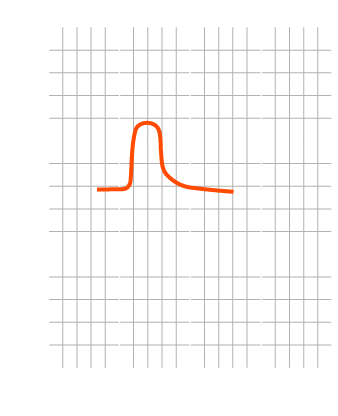
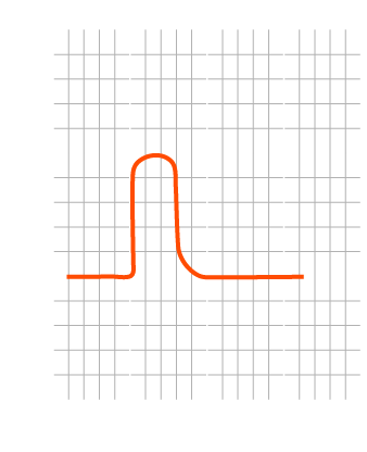
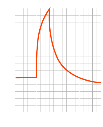
In compliance with design philosophy, the STIMFIT electrical muscle stimulator supply stimulation impulses with a voltage waveform which is very similar to the actual waveform of a motor neuron action potential. An extremely important fact is that such wave form does not undergo any modification or distortion whatsoever while passing through the tissues, thus reaching motor nerves with a form which is very similar to the biological one. This is also due to the presence of HI frequency components (up to 1 MHz), which is deep tissue penetration with less difficulty when compared to any other impulse waveform.
Action Potential
brain signal
STIMFIT
During treatment
Conventional EMS
During treatment
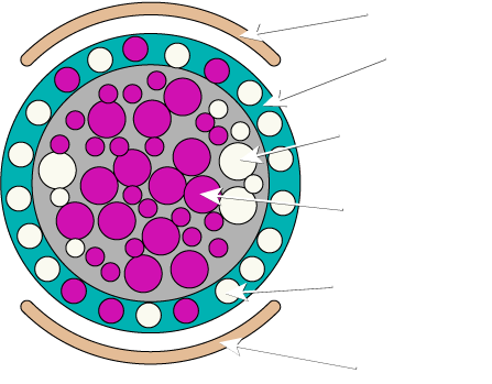
Needless to say, the STIMFIT impulse waveform – so similar to neural physiology – is easily recognized by the neuromuscular system. This implies lower stimulation thresholds and allows to combine the recruitment of a much larger amount of muscle fibers with a lower quantity of electricity and an unequaled level of skin comfort.
STIMFIT Muscular Recruitment
Deeper stimulation
Less electric energy wasted through the skin
More skin comfort
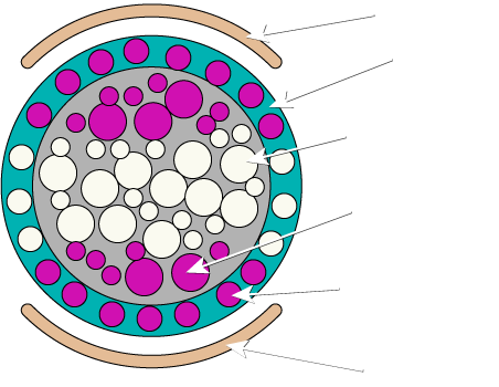
Conventional EMS Muscular Recruitment
Superficial stimulation
More electric energy wasted through the skin
Less skin comfort
7. Skin Pain Awareness Feeling
Pain receptors in the skin are activated by external stimuli. Nerves carrying pain signals from such receptors differ slightly from motor neurons by diameter and conduction speed. Nevertheless, if stimulation energy is powerful enough and if their threshold to a particular stimulation waveform is exceeded, they may be activated by the electric impulses from the stimulator.
The goal of effective stimulation is to maximize its effects on the muscle without any pain on the skin surface. This is now possible thanks to waveform features of the STIMFIT device and to the low voltage of its impulses. Unlike other device, the STIMFIT is pain-free and, a rather pleasant experience as no pain receptor is activated by its waveform.
If the stimulation is done at maximal intensity and for extended time it could result in latent muscular pain due to intense exercise this is the healthy sign proving that the stimulation session was effectively equivalent to physical training.
